From the Eyes to the Brain
To this point in our discussion of the visual system, we’ve examined the eyeball and particularly the layers of cells at the back of it that comprise the retina, which we told you was really an outgrowth of the brain. It’s now time to consider where the information about the visual scene that the receptors have changed into nerve impulses goes in the brain. What are the pathways taken by this visual information, and what brain structures are involved?
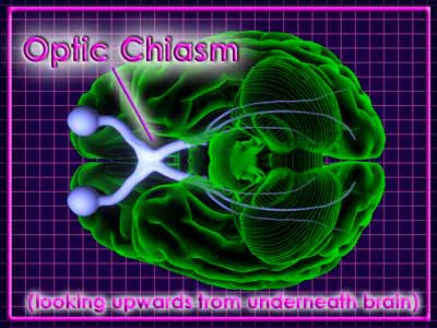
As we told you earlier, the receptor cells (rods and cones) are connected to bipolar cells, which in turn are connected to ganglion cells. The axons of the ganglion cells make up the optic nerve, which leaves the back of the eye at the optic disk, also known as the blind spot because there are no receptors there. The figure below shows the major structures and pathways of the visual system.
As you can see, the optic nerve leaves the back of each eyeball. The optic nerves from your two eyes converge under your brain to form an “X-shaped” structure called the optic chiasm. Appropriately, the Greek letter “chi” is an X.